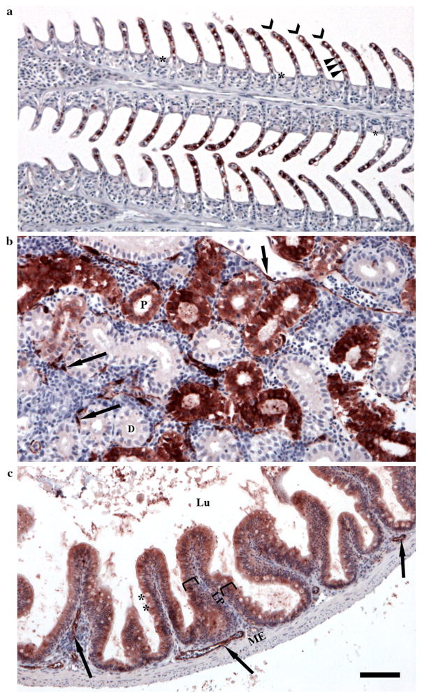
Fig. 1.
Scoring system for determining localization and abundance of CYP1A protein (red staining) in gills, head kidney, and intestine from adult Gulf killifish (Fundulus grandis). a Gills were scored according to degree of staining in pillar cells (arrow heads) along the lamellae (chevrons), and for the combined degree of staining in other remaining cells including mucus cells within the interlamellar region (asterisks), vascular endothelial cells (not identified in a, see b and c for examples of vascular endothelial cells), and epithelial cells covering the surface of the gill and the interlamellar region. b Head kidneys were scored for the staining of the epithelial cells of the distal and proximal tubules (D and P) and for the degree of staining in vascular endothelial cells (arrows). c Intestinal tissues were scored based on degree of staining in the epithelial and mucus cells (asterisks) of the mucosal layer (brackets) and for the degree of staining of vascular endothelial cells. Lu lumen, LP lamina propria, ME muscularis externa. See Table 3 for additional information. All tissues sectioned at 4 μm and imaged with a 20X objective. Scale bar = 50 μm. All slides were counterstained with hematoxylin (blue)

