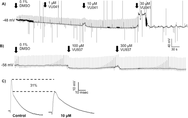
Figure 8.
Recordings of the electrically-evoked EPSPs at the neuromuscular junction in D. melanogaster larvae after exposure to VU041. (A) Representative time course of increasing VU041 concentrations applied to the body wall musculature while recording evoked EPSPs. The remaining transients after block of the EPSP at 30 μM are stimulus artifacts, which are also reflected by any negative excursions from baseline in all traces (artifact amplitudes were truncated from the recordings for clarity of display). The increase in membrane potential after the application of 30 μM is an artifact from the application of the drug and is not a direct response to VU041 since it was not observed in any other recording. (B) Representative time course of increasing VU937 concentrations applied to the body wall musculature while recording evoked EPSPs. (C) Representative evoked EPSP waveforms after exposure to 10 μM VU041 when compared to control.

