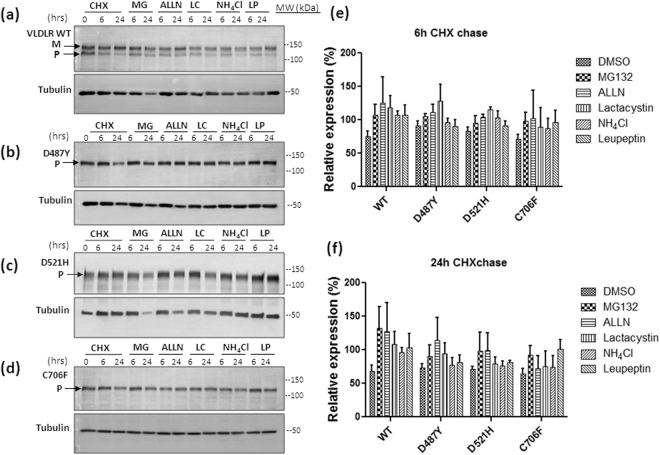
Figure 3.
Effect of proteasomal or lysosomal inhibitors on the degradation rates of VLDLR WT and mutants. HEK-293 cells transiently expressing the VLDLR WT (a) or mutants (b–d) mutants were treated with cycloheximide (CHX) in the presence of either inhibitors of proteasome (10 µM MG132, 10 µM ALLN or 10 µM lactacystin) or lysosome (0.1 mM leupeptin or 20 mM NH4Cl) as indicated. Total cell lysates were analyzed by immunoblotting against HA. Relative amounts of respective proteins remaining at the indicated time points were quantified, and normalized to tubulin levels. Tubulin normalized VLDLR protein levels at 0 h were defined as 1.0 for each panel. The experiments were performed thrice with identical results. (e) Graph representing the relative mean densities of vehicle (DMSO) or inhibitor treated wild type VLDLR and all the mutants at 6 h time-point (f) Graph representing the relative mean densities of DMSO or inhibitor treated VLDLR wild type and all the mutants at 24 h time-point. Error bars represent SEM from n = 3 independent experiments. Regions cropped from separate images are demarcated with borders. Unprocessed original scans of western blots are shown in Supplementary Figure S10.

