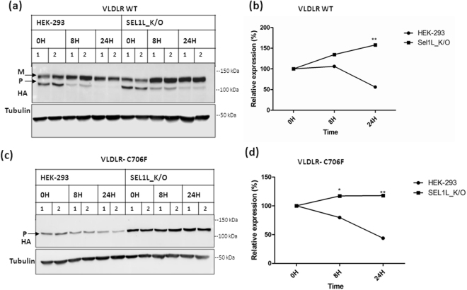
Figure 6.
Delayed degradation of VLDLR WT and mutant in SEL1L knockout cells. VLDLR WT or the mutant C706F was transfected in HEK-293 cell lines and SEL1L Knockout (K/O) cell lines (generated by gRNA1) in parallel. At 24 h after transfection, the cells were incubated with 100 µg/ml CHX for 24 h and collected at different times (8 h & 24 h) for western blot. Replicates for each time point were analyzed in the same blot. The blots were probed against HA and tubulin. Tubulin normalized VLDLR protein levels at 0 h were defined as 100%. (a) Immunoblots showing the turn-over of VLDLR-WT protein. (b) Densitometric analysis of three independent experiments are reported in the graph. (*) p ≤ 0.05; (**); p ≤ 0.01; (***) p ≤ 0.001; Student’s t-test. (c) Turn-over rates of VLDLR C706F in HEK-293 cells and SEL1L knock-out cell lines. (d) Graph representing densitometric analysis of three independent experiments. p values as indicated in (b). Regions cropped from separate images are demarcated with borders. Unprocessed original scans of western blots are shown in Supplementary Figure S13.

