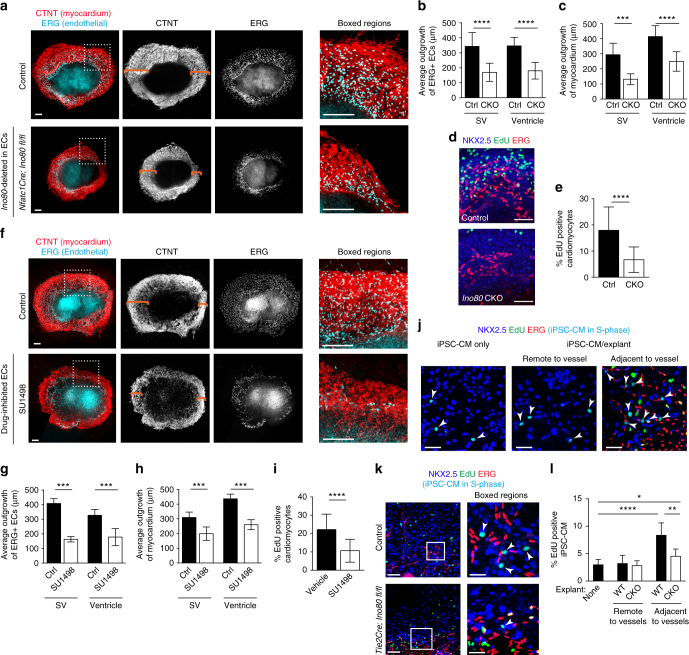
Fig. 6.
Endothelial cells support myocardial growth in the absence of blood flow in an Ino80-dependent manner. Heart ventricle and sinus venosus (SV) explants cultured for 5 days and immunostained for myocardium and endothelial cells. a Confocal images of ventricular explants show that expansion of myocardium (orange brackets) and endocardial-derived cells is decreased when Ino80 is deleted using Nfatc1Cre. Images are representative of the following number of replicates: control, n = 8 heart explants; mutant, n = 5 heart explants. Scale bars: 100 μm (low and high magnification). b–e Quantifications show that endothelial sprouting (b), myocardial outgrowth (control, n = 8 heart explants; mutant, n = 5 heart explants) (c) and cardiomyocyte proliferation (control, n = 6 heart explants; mutant, n = 8 heart explants) (d, e) are significantly reduced. Images are representative of the following number of replicates: control, n = 6 heart explants; mutant, n = 8 heart explants. Scale bars: 100 μm (d). Error bars in graphs are standard deviation. ***P < 0.001; ****P < 0.0001, evaluated by Student’s t-test (d). f–i Inhibiting endothelial cell growth through targeting VEGFR with SU1498 recapitulates the effect of Ino80 mutation (f), significantly decreasing vessels sprouting (g), myocardial expansion (control, n = 6 heart explants; SU 1498 treatment, n = 6 heart explants) (h), and cardiomyocyte proliferation (control, n = 10 heart explants; SU 1498 treatment, n = 8 heart explants) (i). Error bars in graphs are standard deviation. ***P < 0.001; ****P < 0.0001, evaluated by Student’s t-test. Scale bars: 100 μm (low and high magnification). Images are representative of the following number of replicates: control, n = 6 heart explants; SU 1498 treatment, n = 6 heart explants (f). j, k Images of human induced pluripotent cell-derived cardiomyocytes (iPSC-CMs) cultured either alone or with mouse ventricle explants. Proliferating NKX2.5+EdU+ iPSC-CMs are turquoise and indicated by arrowheads. Images are representative of the following number of replicates: control n = 10 heart explants, mutant n = 4 heart explants, iPSC-CM only control n = 7. Scale bar: 25 μm (j); 100 μm (k; low magnification) and 25 μm (k; high magnification). l Quantification of iPSC-CM proliferation within three cells lengths from endothelial cells reveals a significant increase over regions distant from vessels or iPSC-CM cultures only (control n = 10 heart explants; mutant n = 4 heart explants; iPSC-CM only control n = 7). This increase is significantly dampened when endothelial cells are depleted of Ino80. Error bars in graphs are standard deviation. *P < 0.05; **P < 0.01; ****P < 0.0001, evaluated by Student’s t-test. Scale bars: 100 μm

