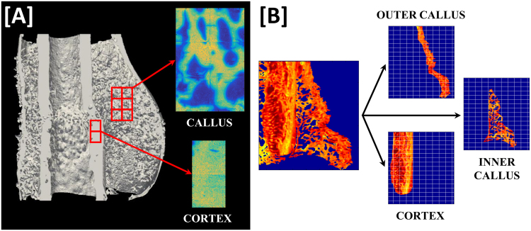
Figure 1.
Illustration of the regions of interest for characterization of fracture site composition and nano-structure. (A) FTIR spectra were acquired of the depicted inner callus and cortical regions. (B) Identical regions in all samples of approximately 5 × 4 mm were scanned with the SAXS/WAXS setups. Measured fracture sites were then sub-divided into inner callus, outer callus and cortical regions of interest as illustrated and parameters for each sample were evaluated within these regions of interest.

