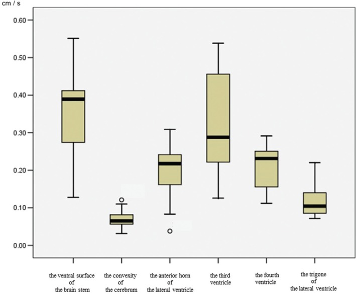
Fig. 1.
Box plots of CSF velocity using 4D-PC in each region. There is markedly increased CSF velocity in the ventral surface of the brainstem. In the ventricular system, the CSF velocity in the third ventricle is increased, followed by the fourth ventricle and the anterior horn of the lateral ventricle. The CSF velocity in the trigone of the lateral ventricle is markedly lower than in other regions. The CSF velocity in the convexity of the cerebrum is also lower than that of the ventral surface of the brainstem. Outside values are indicated by a small “0”.

