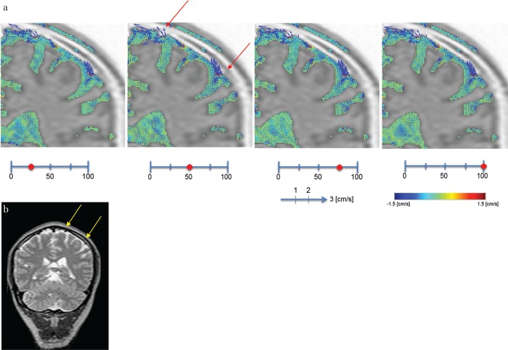
Fig. 3.
Illustrative case of a 54-year-old man. (a) Increased magnification of CSF velocity mapping by the 4D-PC method. A limited increase of CSF velocity around the vessels of the convexity of the subarachnoid space is shown (arrows), while slow CSF velocity in the trigone of the lateral ventricle is also shown. (b) The T2-weighted image of the same volunteer as (a) shows a vascular structure in the convexity of the cerebrum (arrows).

