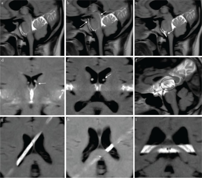
Fig. 5.
The t-SLIP images in various parts of the intracranial CSF cavity (a, b, c) 28-year-old volunteer. The tag was applied at the perpendicular axis of the brainstem. The labelled CSF moves in both upper and lower directions at the ventral surface of the brainstem and Sylvian aqueduct (arrow). (d) 28-year-old volunteer. The tag was applied at the third ventricle in the coronal image. The CSF from the third ventricle surges up into the anterior horn through the FOM (arrow). (e) 56-year-old volunteer. The tag was applied at the third ventricle in the axial image. The labeled CSF from the third ventricle surges up into the anterior horn through the FOM (arrow). (f) 56-year-old volunteer. The tag was applied at the third ventricle in the sagittal image. Marked turbulent CSF motion is observed in the third ventricle (circle). (g) 28-year-old volunteer. The tag was applied above the CP. No obvious CSF movement around the CP is seen. (h) 28-year-old volunteer. The tag was applied perpendicular to the axis of the CP. No CSF upward or downward movement is noted in the trigone. (i) 28-year-old volunteer. The tag was applied at the trigone. No turbulent CSF motion is noted in the trigone.

