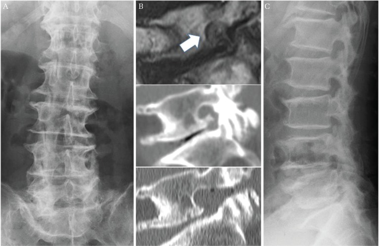
Fig. 4.
A representative case with excellent outcome in group 2. A 74-year-old man underwent a lateral foraminotomy for right L5/S LFS. His preoperative radiograph showed 0.4° of disc wedging at L5/S concave to the right (A), and 35.8° of lumbar lordosis (not shown). His MRI and CT demonstrated cephalocaudal stenosis of the right neural foramen at L5/S, and sufficient decompression was achieved after surgery (B) (upper; preoperative MRI sagittal image, middle; preoperative CT sagittal image, lower; postoperative CT sagittal image, white arrow demonstrates narrowed neural foramen). 18 months after surgery, he showed good clinical outcome with no significant deterioration of lumbar alignment. His radiograph at final follow-up showed 0.5° of disc wedging at L5/S concave to the right (not shown), and 37.4° of lumbar lordosis (C). LFS: lumbar foraminal stenosis, MRI: magnetic resonance imaging, CT: computed tomography.

