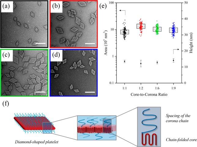
Figure 1.
TEM micrographs of (a) PLLA36-b-PDMAEMA315, (b) PLLA36-b-PDMAEMA216, (c) PLLA36-b-PDMAEMA57, and (d) PLLA36-b-PDMAEMA35 diamond platelets. (e) Jitter box plot and average height data showing the negligible difference in area (as determined by TEM) and height of diamond platelets (as determined by AFM) regardless of block ratio. Samples were self-assembled at 90 °C for 4 h, cooled to room temperature, and aged for 1 day. Samples were stained with uranyl acetate. Scale bar = 1 μm. (f) Schematic of a crystallized PLLA-b-PDMAEMA polymer chain within a diamond-shaped platelet, showing a representation of the chain-folded core block and the spacing of the corona chain.

