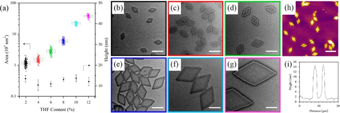
Figure 2.
(a) Jitter box plot and average height data showing the exponential increase in PLLA36-b-PDMAEMA216 diamond nanoplatelet area (as determined by TEM) with increasing THF content with a negligible difference in height (as determined by AFM). TEM micrographs of nanoplatelets prepared in ethanol with (b) 2%, (c) 4%, (d) 6%, (e) 8%, (f) 10%, and (g) 12% THF. Scale bar = 1 μm. (h) AFM image and (i) height profile of nanoplatelets prepared with 12% THF. Scale bar = 5 μm. Samples were self-assembled at 90 °C for 4 h and cooled to room temperature. Note that no change in size was observed on prolonged heating of the solutions (Figure S9).

