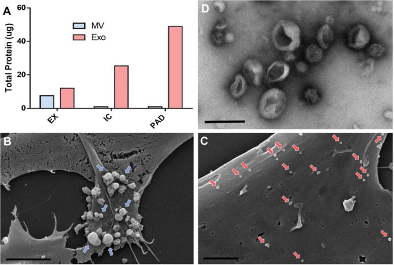
Figure 3.

Mesenchymal stem cells (MSCs) increase secretion of exosomes upon exposure to PAD-like conditions. (A): Quantification of total protein content of vesicles derived from MSC under EX, IC, and PAD culture conditions using DC assay. (B): Scanning electron micrograph of MSCs cultured in EX culture conditions indicating microvesicle release (blue arrows) from the cell surface (scale bar = 5 μm, × 5k). (C): Scanning electron micrograph of MSCs cultured under PAD conditions (scale bar 2 μm, × 10k) indicating exosome adhesion to cell surface (red arrows). (D): Transmission electron micrograph of MSC derived exosomes with 2% uranyl acetate negative staining (scale bar 200 nm, × 25k). Abbreviations: EX, expansion condition; Exo, exosomes; IC, intermediate condition; MV, microvesicle; PAD, peripheral arterial disease.
