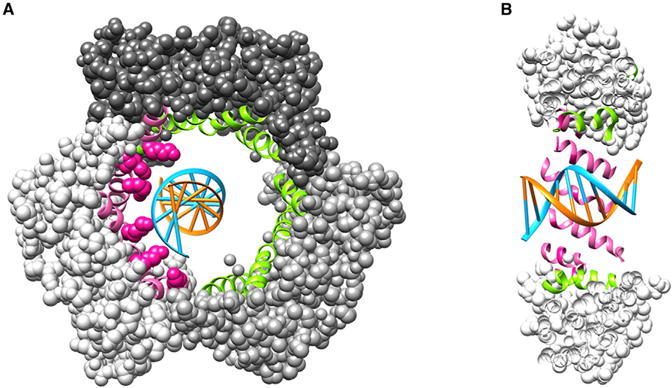
Figure 1. Human PCNA–DNA crystal structure.

(A) Front view of the PCNA trimer–DNA (PDB ID 5L7C) in space-filling representation; each subunit is a different shade of grey. The α helices lining the central chamber are in ribbon representation. In pink are the helices that interact with DNA (the blue strand), and in green are the remaining α helices (the six lysine side chains that interact with DNA are in pink space-filling representation). DNA is in blue and orange. (B) Cut away side-view of PCNA–DNA. The representation and colors of the protein and DNA are the same as in (A).
