
Fig. 7.
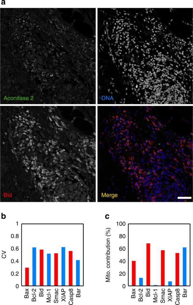
Colon cancer cells show variability in apoptotic proteins and mitochondria. a Colon cancer section stained with Aconitase 2, Bid and DAPI. This image illustrates the variability in expression of Bid and Aconitase 2. Scale bar: 50 μm. b Coefficient of variation (CV) of several proteins involved in the apoptosis pathway. c Mitochondrial contribution to variability of the apoptotic proteins. Data are representative of four independent biopsies. Statistical quantities were obtained from ensembles of 500–1000 cells for each protein antibody