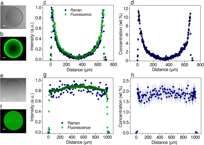
Figure 1.
CRM and CLSM imaging of alginate microbeads with heterogeneous (a–d) and homogeneous (e–h) spatial distribution of alginate. (a,e) CLSM image in the transmission mode. (b,f) CLSM image in the fluorescence emission mode at the equatorial microbead cross-section. (c,g) Overlay of CLSM and CRM intensity profiles at the equatorial microbead cross-section. (d,h) Spatial distribution of alginate in the microbeads shown in (c) and (g), respectively, expressed as the absolute alginate concentration in wt.%. Bars in (a,b,e,f) are equal to 100 μm.

