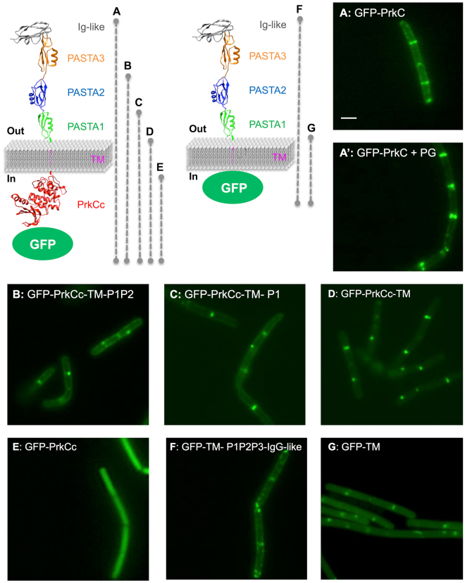
Figure 1.
Localization of full-length and truncated GFP-PrkC in B. subtilis. 3D representation of GFP-PrkC fusion proteins, with A to G letters and dashed lines indicating the length of the protein. PrkC molecular graphic was modeled with the UCSF Chimera package (supported by NIGMS P41-GM103311) from the 3PY9 PDB structure for the extracellular domain and 4EQM PDB structure for the intracellular domain. Strains were grown on LB medium at 37 °C and all the forms of GFP-PrkC proteins were expressed from the Pxyl promoter in the presence of 0.5% xylose and with 3 µg/ml of PG fragments for the full-length protein. PrkC localization was analyzed by fluorescent microscopy for strains: A: SG278 (ΔprkC, amyE::gfp-prkC), A’: SG278 in the presence of PG fragments, B: SG467 (ΔprkC, amyE::gfp-prkCc-TM-P1P2), C: SG466 (ΔprkC, amyE::gfp-prkCc-TM-P1), D: SG465 (ΔprkC, amyE::gfp-prkCc-TM), E: SG355 (ΔprkC, amyE::gfp-prkCc), F: SG497 (ΔprkC, amyE::gfp-TM-P1P2-Ig-like) and G: SG498 (ΔprkC, amyE::gfp-TM). The scale bar for microscopy images represents 2 µm.

