
Fig. 5.
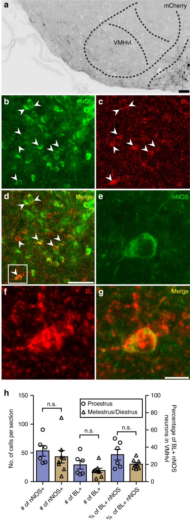
VMHvl nNOS neurons are connected to kisspeptin neurons. a mCherry-immunoreactive projections were detected in the VMHvl after injection of AAV-ChR2 into the RP3V of KissIC/R26-BIZ mice (scale bar = 50 μm); n = 3. b–d Transsynaptic tracing reveals that nNOS neurons in the VMHvl are (either directly or indirectly) connected to kisspeptin neurons (scale bar = 50 μm). BL+ nNOS neurons are indicated with arrows. e–g Zoomed-in image of insert shown in d (scale bar = 10 μm). h The number of neurons expressing nNOS in the VMHvl, the number of BL+ cells, and the overall percentage of BL+ nNOS neurons were not found to be significantly different (Bonferroni’s multiple comparison test) between proestrus and metestrus/diestrus. n = 6 for proestrus, n = 8 for metestrus/diestrus. Bars represent the mean ± SEM. For all experimental details, see Supplementary Table 1