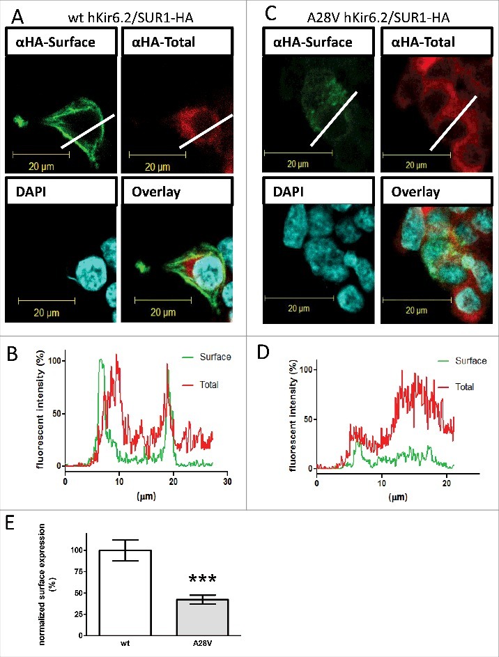
Figure 2.

Surface expression of KATP channels in HEK cells. An HA-tag was inserted into the extracellular domain on rSUR1 and then co-transfected with either wild-type hKir6.2 (A) or A28V hKir6.2. As Kir6.2 must be co-assembled with SUR1 for correct trafficking, the subcellular distribution of HA staining should faithfully represent the KATP channel distribution. KATP channels located on the cell surface were labeled green. Total KATP channels were labeled red, and cell nuclei were counterstained with DAPI (blue). The wild-type KATP channels were clearly visible on the cell surface (A), but A28V hKir6.2-containing channels were not readily observed on the cell surface (C). (B&D): Cross-sectional staining intensity profiles of HEK cells expressing wild-type hKir6.2 (C) and A28V hKir6.2 (D). The profiles were determined from cross-sections indicated by the white lines in A and C. Surface staining signals are clearly visible in HEK cells transfected with wild-type hKir6.2, as the green line shows distinct peaks at the cell boundary. By contrast, in HEK cells transfected with A28V hKir6.2, the cell surface boundary is not clearly demarcated (green line, D). (E) Quantitative analysis of KATP channel surface staining signals. KATP channels containing A28V hKir6.2 had greatly reduced surface staining signals compared to wild-type hKir6.2 containing KATP channels (***p < 0.0005, Mann-Whitney U-test n = 11 for each group).
