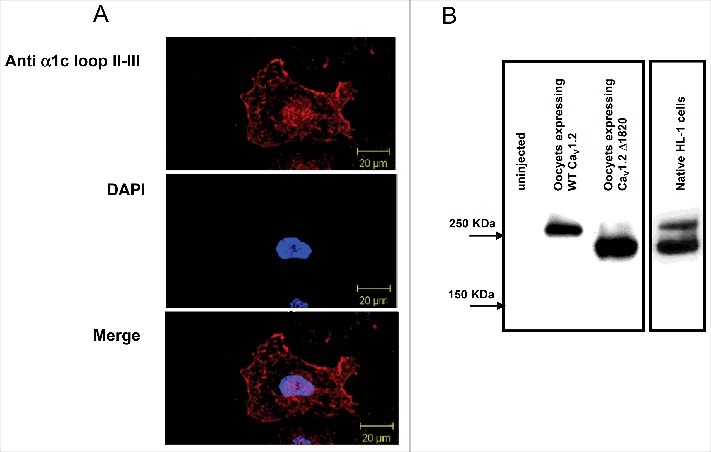
Figure 3.

Native HL-1 cells express α1C. (A) Confocal images of HL-1 cells stained with α1C antibody. (B) Western blot of native HL-1 cells, with lysates of oocytes expressing either full-length of dCT-truncated α1C as a reference. HL-1 cells express 2 distinct molecular weights of α1C, probably corresponding to full-length and dCT-truncated channels.
