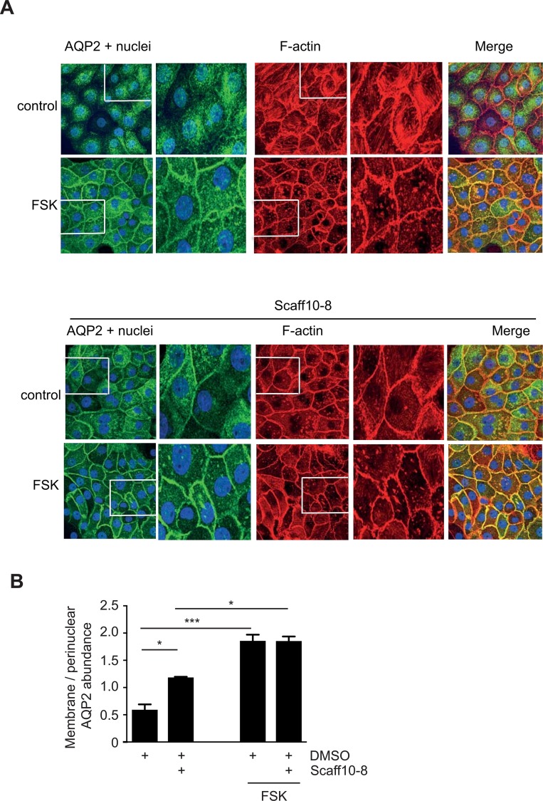
Fig 8. Scaff10-8 promotes the redistribution of AQP2 to the plasma membrane of primary IMCD cells, which is independent of cAMP elevation and associated with depolymerization of F-actin.
(A) Upper panel. IMCD cells were treated with DMSO (1%; control), the solvent of Scaff10-8, or forskolin (10 μM, 30 min) to stimulate cAMP synthesis. Lower panels: The cells were treated for 1 hour with Scaff10-8 (30 μM) alone (control) or with Scaff10-8 and forskolin. Forskolin (10 μM) was added for the final 30 min of Scaff10-8 incubation. AQP2 (green) was detected with specific primary and Cy3-coupled secondary antibodies, F-actin (red) with Alexa Fluor 647-Phalloidin and nuclei with DAPI (blue). Signals were visualized using a laser scanning microscope. Representative images are shown. n = 3. The magnified views were derived from the indicated boxes. (B) The signal intensities arising from intracellular and plasma membrane AQP2 were recorded, related to nuclear signal intensities, and the ratios of plasma membrane to intracellular fluorescence signal intensities were calculated (n = 30 cells per condition). Ratios > 1 indicate a predominant localization of AQP2 at the plasma membrane. Statistically significant differences were calculated using one-way ANOVA with posthoc Bonferroni. Mean ± SEM; *, p ≤ 0.05; *** p ≤ 0.001.

