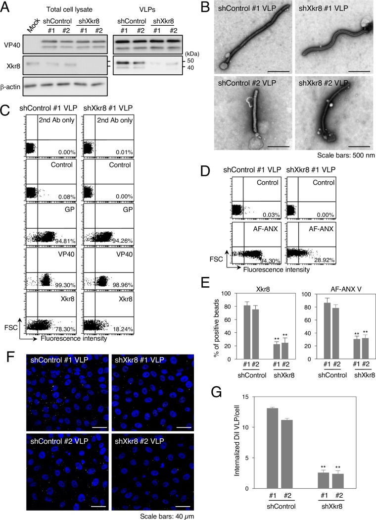
Fig 5. Role of Xkr8 in the externalization of PS on Ebola VLPs and PS-dependent VLP internalization.
(A) Downregulation of Xkr8 by shRNA. Individual 293T clones transduced by shRNA plasmids were transfected with the expression plasmids of EBOV VP40, GP, and NP. At 48 h.p.t., the cells and culture medium were harvested. Viral particles were purified from culture medium by ultracentrifugation. Expression of VP40 and Xkr8 in total cell lysates and VLPs was analyzed by western blotting with rabbit polyclonal antibodies against VP40, Xkr8, or β-actin. (B) Negative staining of VLPs obtained from individual shRNA clones. Individual shRNA cell clones were transfected with the expression plasmids of VP40, GP, and NP. At 48 h.p.t., the culture medium was harvested. Viral particles were purified from the culture medium by ultracentrifugation followed by negative staining. Scale bars; 500 nm. (C–E) The effect of Xkr8 knockdown on the incorporation of viral proteins and Xkr8 into VLPs and on the externalization of PS on the surface of Ebola VLPs. (C) Ebola VLPs obtained from shControl #1 (left) or shXkr8 #1 (right) clones were conjugated with latex beads. The beads were then incubated with rabbit polyclonal antibodies against EBOV GP, VP40, or Xkr8 followed by flow cytometric analysis. 2nd Ab indicates samples that were not treated with primary antibody. As a control, the rabbit anti-LASV GPC polyclonal antibody was used. (D) For detection of externalized PS on the VLPs, the beads were incubated with AF-ANX V and subsequently subjected to flow cytometric analysis. The percentages of the positive populations are indicated. X-axis: fluorescence intensity, Y-axis: forward scatter corner signals. The results are representative of three individual experiments. (E) Summary of the binding of the anti-Xkr8 antibody (left) and AF ANX V (right) to individual shControl VLP- or shXkr8 VLP-conjugated beads. Each experiment was performed in triplicate and the percentages of the positive populations are presented as the mean ± SD. **, P < 0.01 versus respective control (Student’s t test). (F, G) The effect of Xkr8 knockdown on the internalization of VLPs. (F) Purified VLPs from individual shRNA clones were labeled with DiI and adsorbed to Vero-E6 cells for 30 min at room temperature. After incubation for 2 h at 37°C, surface-bound virions were removed by trypsinization and the internalization of Ebola VLPs was analyzed by using a confocal laser scanning microscope. (G) The number of internalized DiI-VLPs in each of 10 individual clones was measured. Each experiment was performed in triplicate and the relative uptake efficiencies are presented as the mean ± SD. **, P < 0.01 versus respective control (Student’s t test).

