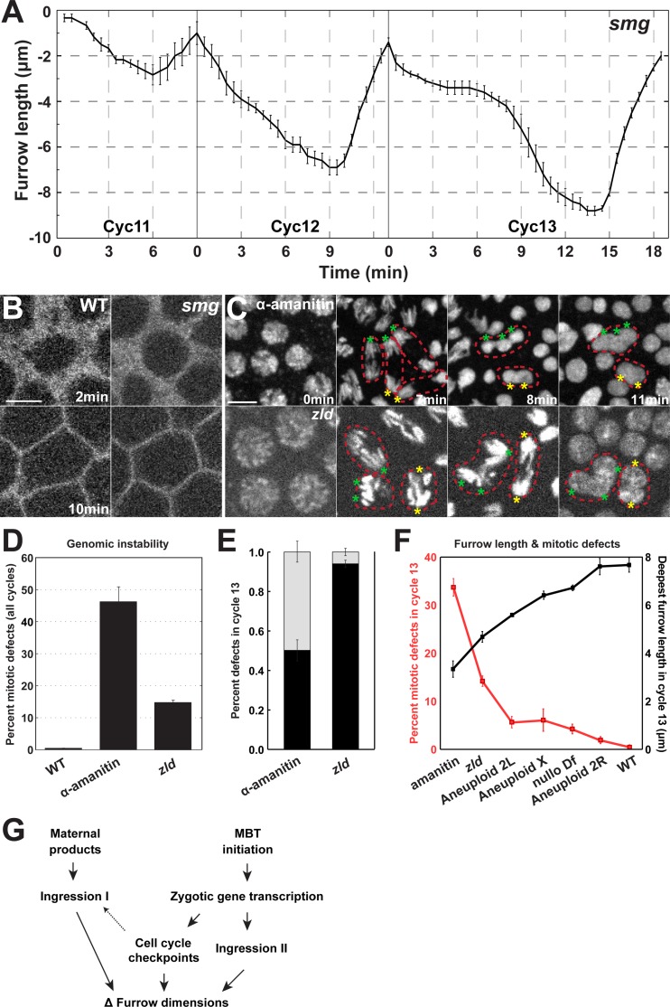
Fig 7. Maternal gene decay is a minor contributor to furrow dynamics.
(A) smg mutant furrow dynamics (cycle 11: n = 3; cycle 12 and 13: n = 5). (B) Furrow morphology in WT and smg mutant embryos at 2 min and 10 min. A region just adjacent to the furrow tips is shown. Scale bar = 5 μm (C) Mitotic defects in α-amanitin injected and zld mutant embryos. Chromosomes are labeled with Histone:GFP. Red, dotted lines highlight individual mitotic figures that fail to properly segregate chromosomes resulting in polyploid nuclei, and asterisks indicate missegregating chromosomal complements. (D) Percent of nuclei that experience mitotic defects in WT, α-amanitin injected, and zld mutant embryos. (E) Percent of adjacent nuclear fusion (black bar) and mitotic nuclear fusion (grey bar) in α-amanitin injected and zld embryos (n = 3) in cycle 13. (F) Furrow length and mitotic defects are inversely correlated. The percentage of mitotic defects at cycle 13 in α-amanitin injected, zld, Aneuploid 2L, Aneuploid X, nullo, Aneuploid 2R, and WT embryos is presented, as well as deepest furrow lengths during metaphase in cycle 13. Intact furrow length and corresponding mitotic defects are measured in S3J Fig (G) Model for developmental regulation of furrow dimensions in the early Drosophila embryo.

