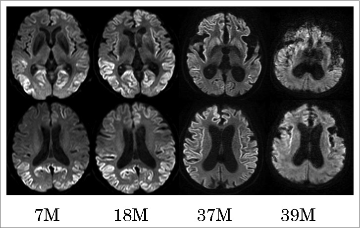
FIGURE 4.

Serial diffusion-weighted images of MR. Serial diffusion-weighted images (DWI) obtained at 7, 18, 37, and 39 months (M) after the onset of initial symptoms. The upper panel shows serial MR images at the level of the basal ganglia, and the lower panel shows those at the slice of corona radiata. Serial DWI showed that cortical hyper-intense area extended across frontal and temporal cortices and bilateral basal ganglia from 7 months to 37 months. DWI signals were diminished in the occipital cortices at 37 months, compared with those at 7 or 18 months, and in the frontal and temporal cortices at 39 months versus 37 months.
