FIGURE 1.

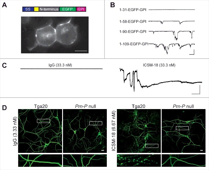
The isolated N-terminal domain of PrPC induces ionic currents, and anti-PrP antibodies induce currents and dendritic degeneration. (A) Top, schematic of PrP(N)-EGFP-GPI constructs containing the N-terminus of PrP fused to EGFP and the PrP GPI attachment sequence. The colored blocks represent the signal sequence (purple), polybasic residues 23–31 (yellow), different portions of the N-terminus (grey), EGFP (green), and the GPI attachment sequence (magenta). Bottom, fluorescence image of N2a cells expressing PrP(1-109)-EGFP-GPI, showing localization of the protein on the cell surface. Scale bar = 10 μm. (B) Representative traces of currents recorded from N2a cells expressing constructs with different lengths of the N-terminus (1–31, 1–58, 1–90 and 1–109). Scale bars: 500 pA, 30 s. (C) Representative traces of currents recorded from cells expressing wild-type PrP in the presence of non-specific IgG or ICSM18. Scale bars: 500 pA, 30 s. (D) Representative images showing dendrite morphology of cultured hippocampal neurons from Tga20 mice (which over-express wild-type PrPC) or Prn-p null mice after treatment for 48 hrs with anti-PrP antibody ICSM18 (6.67 nM) or non-specific IgG (33.3 nM). The cells were stained with an antibody to MAP2 to visualize dendrites. Boxed areas are enlarged below each image. Scale bar = 10 µm.
