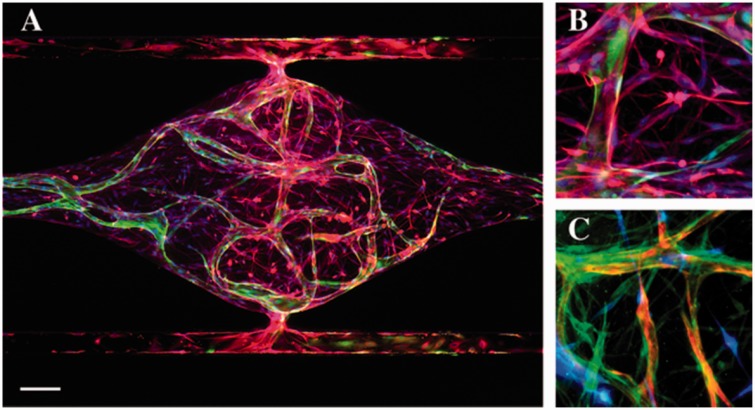
Figure 2.
BBB-on-a-chip supported by living microvascular network. (a) Global view of astrocytes (GFAP – red) and the microvascular network formed by ECFC-EC (green) inside a tissue chamber. Nuclei were stained with DAPI (blue). Scale bar = 100 µm. (b) Astrocytes (GFAP – red) lay down end feet and interact with the vessel (green). (c) Pericytes (PDGFR-β – green) wrap around the vessel (red) and interact with astrocytes (blue)

