Figure 2.
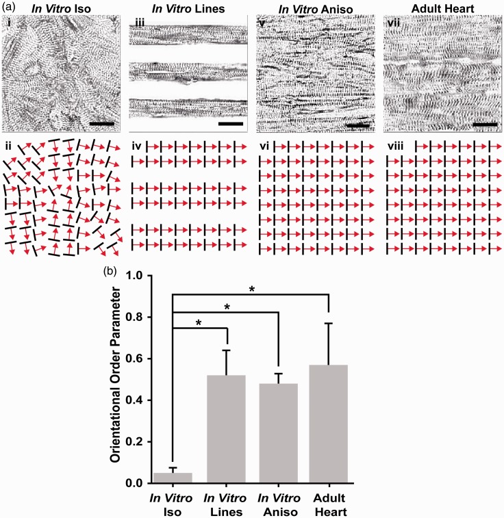
Comparison of global sarcomere alignment in engineered and adult myocardium. (a) Assessment of sarcomeric α-actinin fluorescence micrographs of isotropic samples (i) revealed random z-line organization as illustrated in the schematic below (ii). In contrast, α-actinin micrographs of cardiac myocytes cultured on 15 µm wide FN lines spaced 15 µm apart (iii), displayed the high degree of parallel alignment expected from the ECM patterning, as illustrated in this schematic (iv). The z-lines of cardiomyocytes cultured on 15 µm wide FN lines spaced 2 µm apart (v) also displayed the degree of parallel z-line alignment expected from this ECM pattern, as illustrated in this schematic (vi). Comparison of micro-contact printed engineered myocardium to the z-line organization observed in histological sections of the adult rat ventricular myocardium (vii) reveals a similar level of global sarcomere alignment as illustrated in this schematic (viii). (b) Statistical comparison of global sarcomere alignment quantified using the Orientational Order Parameter revealed that the anisotropic engineered myocardium showed similar levels of alignment to the adult rat ventricular myocardium. n = 3 tissues for In Vitro Iso, In Vitro Lines, and In Vitro Aniso; n = 4 ventricles for Adult Heart. Scale bars = 10 µm. *= P < 0.05 versus In vitro Iso

