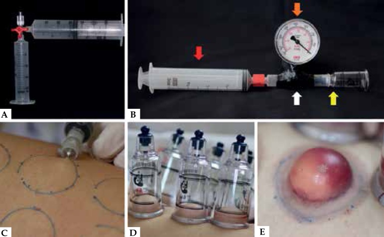
Figure 1.
A - Syringe without plunger connected to the suction syringe through a 3-way connector. B - 2.5 cm suction cup (yellow arrow) connected to a vacuum gauge (orange arrow) adapted to a 5 ml syringe (white arrow), which is then connected to a 60 ml syringe (red arrow). C - Infiltration in two planes in the donor area. D - Suction cups in position (-350 mmHg). E - Subepidermal blister completely formed after 40 minutes

