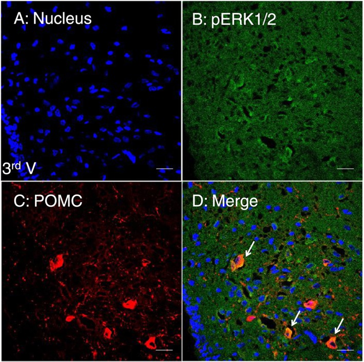
Figure 7.

Double immunofluorescent staining showing the co‐localization of pERK1/2 and POMC proteins in the ARC of amphetamine‐treated rats. Frozen sections of the brain were plated on slides and stained with anti‐pERK1/2 and anti‐POCM antibodies in the hypothalamus. Under the analysis of confocal microscopy, the co‐localization image was seen by the merge of pERK1/2 (green) and POMC (red) immunofluorescent images, giving a yellow colour in the merged image panel. Blue: nucleus. Scale bar: 20 μm. 3rd V: the third ventricle.
