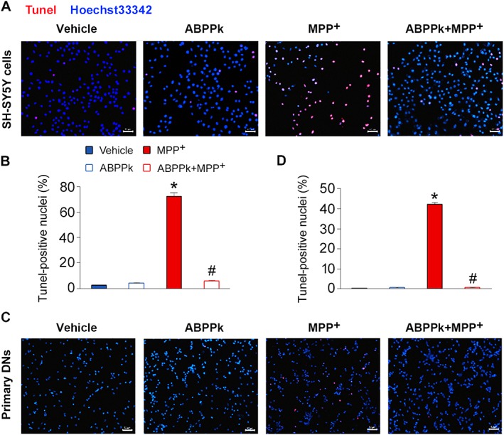
Figure 2.

Tunel assays showing the protective effects of ABPPk. The SH‐SY5Y cells (A, B) and primary dopaminergic neurons (DNs) (C, D) were pretreated with ABPPk at different dosages and exposed to 500 and 50 μM MPP+ respectively. Cell apoptosis was measured by the Tunel assays. Tunel‐positive cells (red) indicate apoptotic cells, and Hoechst33342 was used to stain the nuclei (blue). Scale bar = 50 μm. Error bars are ± SEM. n = 5. *P < 0.05 versus the vehicle treated cells. #P < 0.05 versus the cells treated with MPP+ alone.
