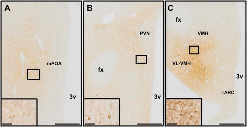
Figure 2.

Images demonstrating the distribution of nNOS-immunoreactive neurones and fibres in the preoptic area (A), paraventricular nucleus (B), rostral arcuate nucleus, ventromedial hypothalamus, and ventrolateral portion of the ventromedial hypothalamus (C). Insets in each panel show higher magnification images of nNOS neurones contained in boxes present in each panel. Scale bars in lower magnification images = 500 μm. Scale bars in higher magnification insets = 50 μm fx, fornix; mPOA, medial preoptic area; PVN, paraventricular nucleus; rARC, rostral arcuate nucleus; VL-VMH, ventrolateral portion of the ventromedial hypothalamus; VMH, ventromedial hypothalamus; 3v, third ventricle.
