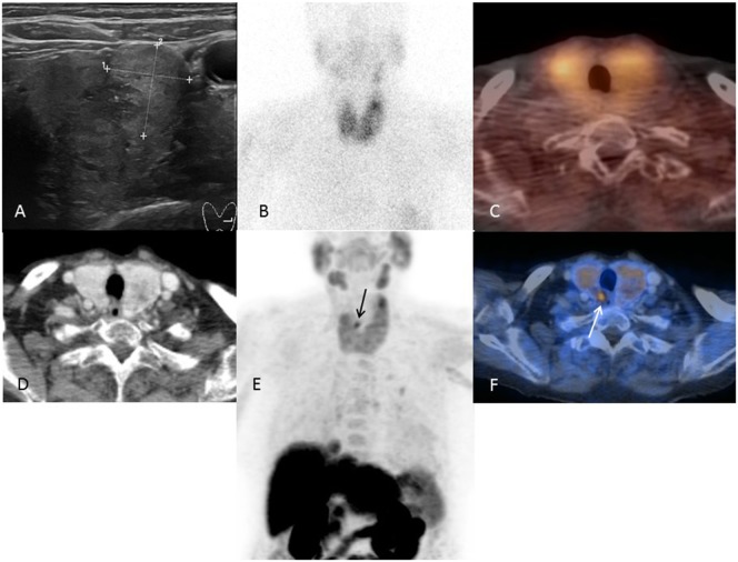
Figure 2.
A representative case of a patient with multinodular goiter and negative conventional imaging. Images of an 82-year-old patient (patient number 9). Ultrasound (A) showing a bilateral multinodular goiter and no visible parathyroid adenoma. 99mTc-sestamibi SPECT/CT (B,C) without detection of a parathyroid adenoma. FCH-PET/CT (D–F) with clear visualization of a small parathyroid retrotracheal adenoma at the upper right pole.

