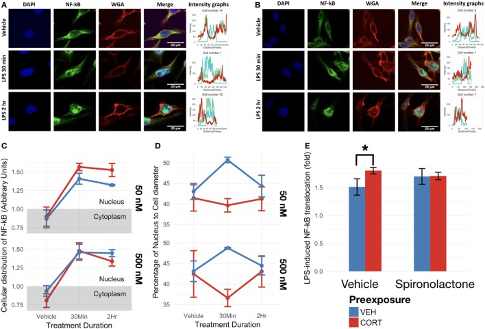
Figure 5.
Corticosterone (CORT) preexposure (50 nM) increased NF-κB translocation, while both 50 and 500 nM prevented increased nucleus:cell diameter 30 min after lipopolysaccharide (LPS) treatment. (A,B) Fluorescent immunocytochemistry for NF-κB (green), nucleus (blue), cell membrane (red), and profile intensity plots across cell diameter shown for one sample cell in vehicle vs 30 min vs 2 h LPS following: (A) vehicle and (B) low concentration (50 nM) CORT preexposure. (C) Summary values representing nuclear expression of NF-κB proportional to total expression of NF-κB across the cell diameter, further normalized to the proportion of nucleus to cell diameter. Value <1: expression distribution favors cytoplasm, value >1: expression distribution favors nucleus. (D) Summary values for nucleus:cell diameter at baseline, 30 min and 2 h following LPS treatment. A lower value reflects decreased nucleus proportion of cell diameter, a measure of increased process length. (E) Effect of mineralocorticoid receptor inhibition via spironolactone treatment on LPS-induced NF-κB translocation (N = 5). All measures were made after preexposure + treatment. Error bars represent mean ± SEM values. Asterisks denote p-values *<0.05, **<0.01, ***<0.001, and ****<0.0001.

