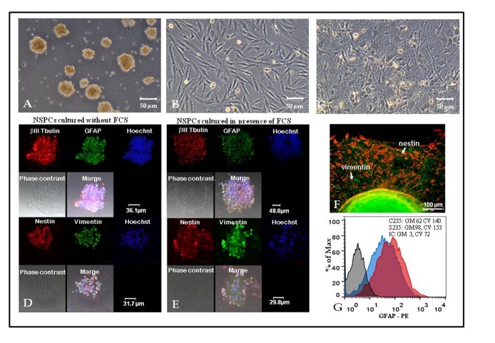
Figure 1.
Neural stem/progenitor cells (NSPCs) isolated and maintained in the presence of fetal calf serum (FCS) demonstrate similarities to “classic” NSPCs grown in serum-free medium.
(A-C) Phase contrast microscopy of S170 culture in FCS-containing medium: neurospheres (A); fibroblast-like cells (B) and web-like structures (C) formed by NSPCs. (D-E) Expression of βIII Tubulin, glial fibrillary acidic protein (GFAP), nestin and vimentin by NSPCs maintained as neurospheres in serum free (D) or FCS-containing (E) medium, confocal microscopy of C235 (serum-free medium) and S235 (FCS-containing medium) cultures. (F) Expression of nestin (red) and vimentin (green) by cells relocating from neurospheres to plastic-adherent state in S170 culture grown in FCS-containing medium, fluorescent microscopy. (G) Flow cytometry analysis of GFAP expression: C235 (serum-free medium) culture— blue curve, S235 (FCS-containing medium) culture— red curve, isotype control (IC) — grey curve. Geometric means (GM) and coefficients of variation (CV) are displayed in the upper right corner.

