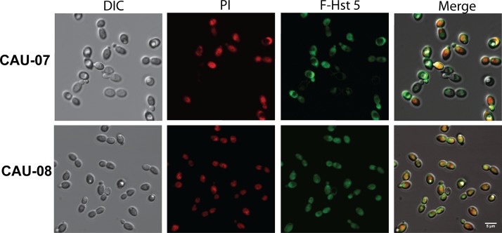
FIG 3.
F-Hst 5 is taken up by C. auris cells. F-Hst 5 (30 μM) and PI (0.5 μM) were added to C. auris cells adherent to a glass coverslip, and intracellular uptake was imaged after 30 min at 23°C using DIC and fluorescence confocal microscopy. Regardless of the killing efficiency of Hst 5, both CAU-07 and CAU-08 transported F-Hst 5 to the cytosol (as visualized by green florescence), resulting in a change in cell topology followed by cell death (as visualized by red PI staining). Bar, 5 μm.

