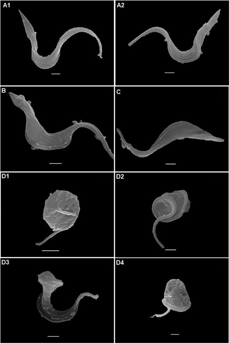
FIG 2.
Scanning electron microscopy examination of T. cruzi bloodstream trypomastigotes (Y strain). (A) Control with no compound exposure. Treatment with 0.88 μM 31DAP069 (B), 0.35 μM 36DAP015 (C), and 10.48 μM 28SMB032 (D) resulted in no morphological alteration for the parasites shown in panel B or C, but the parasites in panels D1 to D4 exhibited body retraction, and those in panels D2 and D3 exhibited a twist of the parasite body. Bars = 1 μm.

