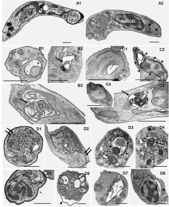
FIG 3.
Ultrastructural effects of AIAs in T. cruzi bloodstream trypomastigotes (Y strain). (A1 and A2) Controls with no compound exposure display the characteristic morphology. Treatment with 0.88 μM 31DAP069 (panels B), 0.35 μM 36DAP015 (panels C), and 10.48 μM 28SMB032 (panels D) resulted in several insults, including dilatation of the flagellar pocket (black star in panels B1, C4, and D6), concentric membranar structures and myelin figures in the cytosol (black arrows in panels B2, B3, C3, and D4), disruption of the Golgi apparatus (asterisk in panels C1 and D7), an endoplasmic reticulum surrounding cytosolic structures (arrowheads in panels C2 and D3), and detachment of the nuclear (short arrow in panel D8) and plasma (short arrow in panel D6) membranes. No alterations were detected in subpellicular microtubules (thin arrows in panel D5) and on the parasite kinetoplast DNA (kDNA) (double arrows in panels D1 and D2). G, Golgi cisternae; M, mitochondria; N, nuclei; F, flagellum. Bars = 500 nm.

