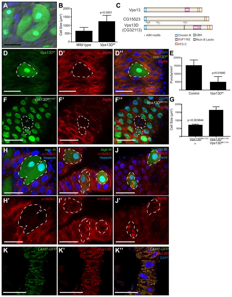
Figure 1. Vps13D functions in programmed cell size reduction and Atg8 puncta formation in the Drosophila intestine.
(A) Control and Vps13D knockdown (green) cells in the Drosophila midgut stained with DAPI (blue).
(B) Quantitation of control wild type and Vps13D knockdown cell size from at least 76 cells in at least 9 intestines of either control or knockdown cell clones.
(C) Schematic of the Vps13 family. Drosophila Vps13D is the only Vps13 family member with a UBA domain. Atg8 interacting motifs analyzed in the manuscript are marked with arrows.
(D-D”) Clonal knockdown of Vpsl3D (green cells) impairs mCherry-Atg8a puncta formation (red).
(E) Quantitation of mCherry-Atg8a puncta in control and Vpsl3D knockdown cell clones from at least 28 clones in 4 intestines.
(F-F”) MiMIC insertion Vpsl3D mutant cell clones phenocopy Vpsl3D knockdown failure in cell size reduction and mCherry-Atg8a autophagy reporter puncta formation (red). The mutant clone (-/-; top cell) and an example of a heterozygous control cell (+/-; bottom cell) are outlined in white.
(G) Quantitation of Vpsl3D mutant and heterozygous control (green) cell size from clones from three intestines.
(H-J') (H-H'), Atg8a (I-I'), and Vpsl3D (J-J') knockdown (green) cells in the Drosophila midgut stained with DAPI (blue) and ubiquitin (fk2) antibody (red). Results are representative of at least three intestines per genotype.
(K-K”) Intestines expressing LAMP-GFP (green) were dissected 2h after puparium formation and stained with DAPI (blue) and Vpsl3D antibody (red). Results are representative of at least three intestines. Scale bars in all images represent 50um.
See also: Figures SI and S2, Table SI

