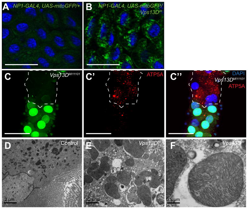
Figure 2. Vpsl3D function is required for mitochondrial clearance and size control in the Drosophila intestine.
(A) Mito-GFP in control gut and (B) Vps13D knockdown midguts 2 h after puparium formation. Results are representative of at least three biological replicates. Scale bars represent 50 μm.
(C-C”) MiMIC insertion Vps13D mutant midgut cells (lacking GFP) possess persistent mitochondrial ATP5A protein compared to neighboring control cells (GFP-positive) indicating a defect in the clearance of mitochondria. Scale bars represent 50 μm.
(D-F) Knockdown of Vps13D results in enlarged midgut mitochondria compared to mitochondria from control w1118 animal midguts. Results are representative of at least three biological replicates.
See also: Figure S3

