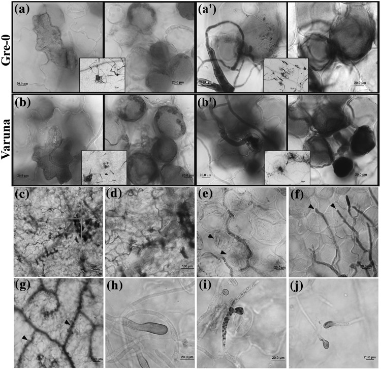
Fig. 2.
Cellular responses to the Alternaria brassicae infection on Gre-0 and Varuna. Representative images of infected Gre-0 and Varuna leaves stained with 3,3'-diaminobenzidine (DAB) to detect hydrogen peroxide (H2O2) accumulation (reddish brown) and trypan blue to detect cell death (blue). At 24 hpi, H2O2 accumulates in epidermal and mesophyll cells at the site of fungal penetration, whereas cell death occurs only in mesophyll both in Gre-0 (a, a′) and Varuna (b, b′). The insets in a, b, a′ and b′ shows localization of DAB and cell death respectively around the penetrated sites in Gre-0 and Varuna at a lower magnification. Infected Varuna leaves showing cell death (CD) following invasion by the fungal hyphae into the healthy mesophyll cells-96 hpi (c, d). Slight coloration of the mesophyll cells (arrowheads) indicates the initiation of cell death around the fungus at 96 hpi (e). Leading edge of the invading hyphae grows into the intercellular space between the mesophyll cells without causing any visible cell death (arrowheads)-96 hpi (f). The extensive spread of cell death observed in Varuna leaf veins (arrowheads) at 7 dpi (g). H2O2 accumulation was observed at fungal hyphal tip and ALS on Varuna (h, i) and Gre-0 (j)

