Figure 2.

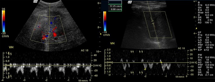
Pulsed Doppler sonographic images show biphasic morphology of the hepatic vein (HV) waveform in an obese dog (left) and triphasic morphology of the HV waveform in an ideal dog (right). VH: hepatic vein.

Pulsed Doppler sonographic images show biphasic morphology of the hepatic vein (HV) waveform in an obese dog (left) and triphasic morphology of the HV waveform in an ideal dog (right). VH: hepatic vein.