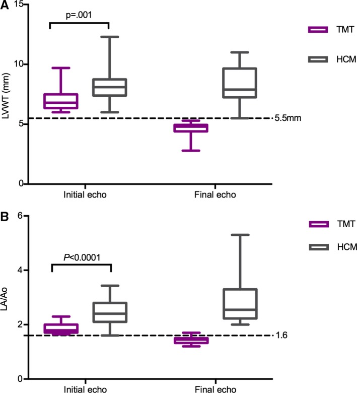
Figure 1.

Left ventricular wall thickness (LVWT) (A) and left atrial size (LA/Ao) (B) in cats with TMT and HCM at presentation and final echocardiographic examination. At presentation, left ventricular walls were thicker in cats with HCM. The left atrium was larger in cats with HCM at presentation and remained dilated over time, while it decreased over time in the TMT population. By definition, the LVWT and LA/Ao decreased between the initial and the final echo in the TMT population, and so those two datasets were not subjected to statistical analysis. Echo, echocardiogram; TMT, transient myocardial thickening; HCM, hypertrophic cardiomyopathy.
