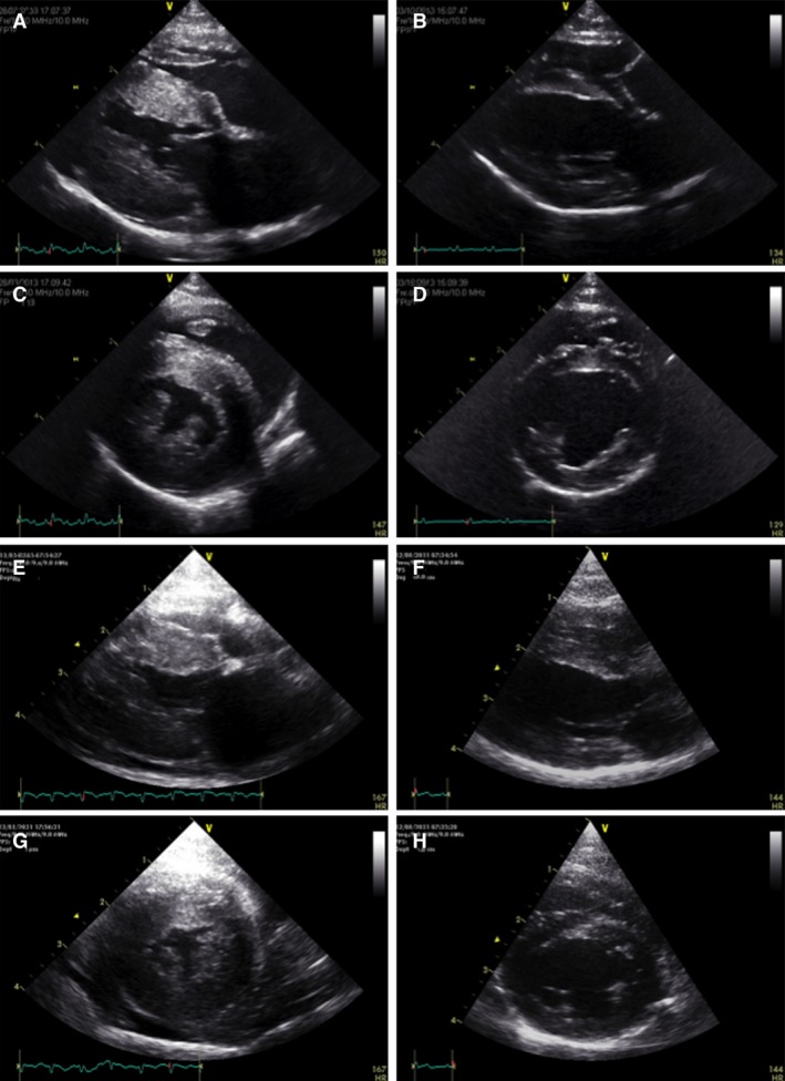
Figure 2.

Right parasternal long‐axis (A, B, E, F) and short‐axis views (C, D, G, H) at end‐diastolic frame from 2 TMT cases at initial presentation (A, C and E, G) and 7 months later (B, D and F, H). The initial severely increased left ventricular wall thickness (A, C, E, G) and mild pericardial effusion (E, G) resolved completely, with a morphologically normal heart 7 months later.
