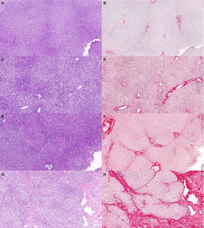
Figure 4.

Hepatic fibrosis in dogs (hematoxylin and eosin: A, C, E, G and picrosirius red: B, D, F, H). Liver sections from dogs with various stages of fibrosis. Note the collagen fibers are more distinct when serial sections are stained with picrosirius red. A, B: absent/minimal fibrosis; C, D: moderate fibrosis with fibrous expansion of the portal tracts; E, F: marked fibrosis with portal‐portal bridging; G, H: very marked fibrosis with discrete nodule formation.
