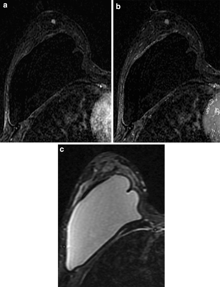
Fig. 11.
Focus with the absence of high signal on T2 sequence. a Postcontrast subtraction T1-weighted image shows a unique 0.4-cm focus with washout delayed kinetics (b) and the absence of high signal on fat-saturated T2-weighted image (c). Because this focus was new, it was assessed as probably benign, BI-RADS 3. Follow-up examination 6 months later showed increase in size of the focus; therefore, biopsy was recommended. MRI-guided wire localization was performed of this focus and surgery yielded invasive ductal carcinoma. For foci with washout kinetics and the absence of high T2 signal, biopsy should be considered

