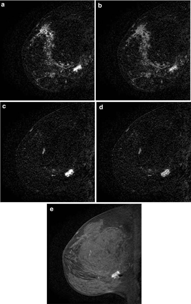
Fig. 13.
Missed MRI-guided biopsy with follow-up demonstrating cancer. a Postcontrast subtraction T1-weighted image shows a 1.2-cm non-mass enhancement (NME) with focal distribution, heterogeneous internal enhancement, and b washout kinetics (arrow), which was suspicious and assessed as BI-RADS 4. MRI-guided biopsy was performed yielding fibrocystic changes and a 6-month follow-up MRI was recommended. At 6-month follow-up, c postcontrast subtraction T1-weighted image shows persistence of the NME and washout kinetics (d). Postcontrast T1-weighted image (e) shows that the susceptibility artifact from the biopsy marker clip is located posterior to the focal NME, which was unchanged in size and appearance suggesting that the NME was not biopsied. Surgical excision yielded carcinoma in situ

