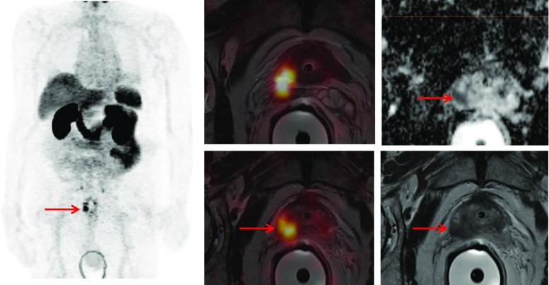
Fig. 1.
PET/MRI demonstrating the primary PC (PSA 16 ng/ml, GS 3 + 4) in the right prostate lobe (red arrow) invading the seminal glands with markedly increased 68Ga-PSMA uptake. The tumor presents with restricted diffusion on apparent diffusion coefficient (ADC) mapping and is hypointense on T2-weighted MRI

