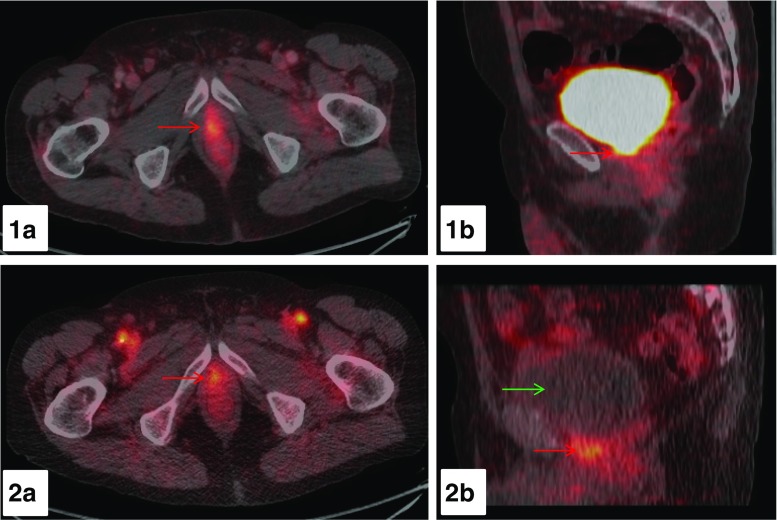
Fig. 3.
68Ga-PSMA-11 PET/CT images of a 72-year-old PC-patient with BR after RP (PSA 4.26 ng/ml). Early dynamic imaging of the pelvis over the first 8 min p.i. and a whole-body scan at 60-min p.i. were performed. At 60-min p.i., a clear distinction between urinary activity within the neck of the urinary bladder and local recurrence is not possible as presented on axial (1a) and sagittal (1b) fused PET/CT images (red arrow). In contrast, on the axial and sagittal fused PET/CT-images (2a, 2b) at 4 min p.i. of the early dynamic PET-acquisition a focal tracer accumulation with a SUVmax value of 4.45 adjacent to the urinary bladder is visible (red arrow) with no tracer uptake in the urinary bladder present (green arrow) consistent with local recurrence

