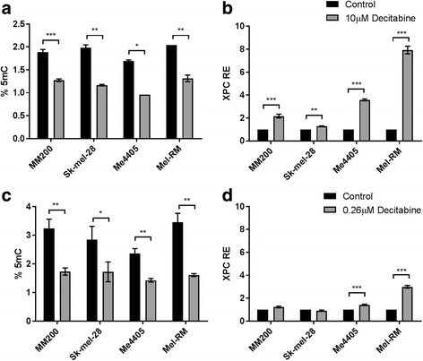
Fig. 1.

Global methylation levels and XPC expression in melanoma after decitabine treatment. Melanoma cell lines were treated with 10 μM decitabine (a) or 0.26 μM decitabine (c) (grey) for 72 h and global methylation levels (%5mC) were quantified and compared to untreated cells (control, black). XPC transcript expression (RE) after 10 μM decitabine (b) and 0.26 μM decitabine (d) was quantified by qPCR and normalised to control. Data represent mean of triplicate experiment, bars = SEM. *p < 0.05, **p < 0.01, ***p < 0.001
