Fig. 2.

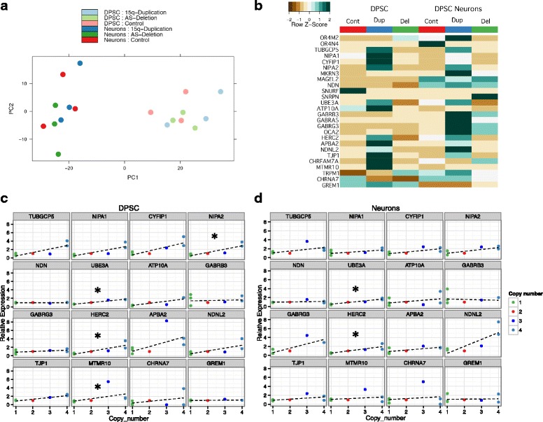
Gene expression changes across the 15q region. a principle component analysis. Note that the main differentiating feature of the gene expression data is the cell type (i.e., DPSC or DPSC-neuron) and not the genotype of the line. b Heatmap of gene expression for genes between BP1-BP3 on 15q11.2-q13.1. Note that some, but not all, genes show elevated expression in the DPSC and DPSC neuronal cultures from duplication lines. The expression differences are less striking for AS deletion lines where only a few specific genes appear significantly downregulated in DPSC and fewer still in the DPSC-neurons. c Graphs of copy number vs gene expression for DPSC in BP2-BP3 protein coding genes. Green dots are AS deletion, red dots are typical control, blue dot is interstitial Dup15q, and light blue dots are idic(15). Note that only NIPA2, UBE3A, HERC2, and MTMR10 showed a significant correlation between copy number and gene expression. Star indicates that these genes showed a significant trend using Jonckheere’s trend test. d Graphs of copy number vs gene expression in neurons for the BP2-BP3 region. Note that only UBE3A and HERC2 still show a significant trend test
